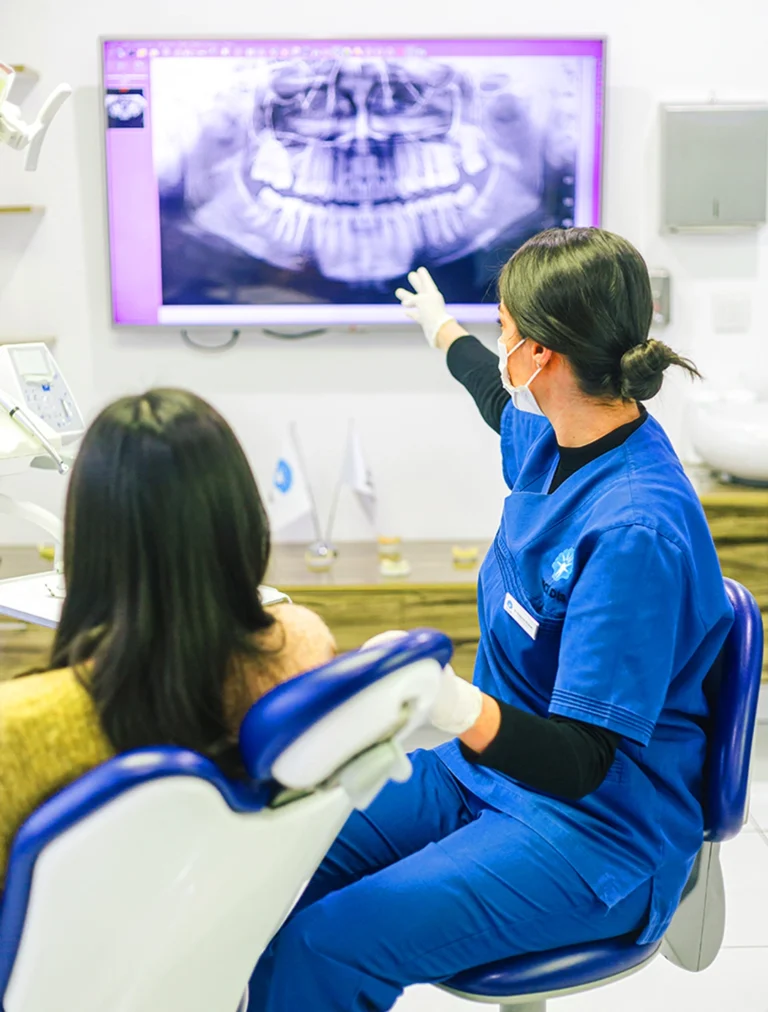
Root resorption can be defined as the melting of the tooth root or shortening of the root length. Root resorption in deciduous teeth that occurs during the change of deciduous teeth and eruption of permanent teeth is physiological. Resorption of deciduous or permanent teeth due to infection or trauma is pathological.
The forces applied to the teeth during orthodontic treatment may cause root resorption for some reasons. This situation can be considered as an undesirable effect of orthodontic treatment in which we make aesthetic and functional corrections in teeth. Biological or mechanical factors alone or in combination can cause this situation. As biological factors, we can list conditions such as gender, age, nutritional diversity, systemic factors, bad habits, trauma history, root canal treatment history and predisposition due to the anatomy of the tooth.
As for mechanical factors, we can say the amount and type of force generated during treatment and the types of apparatus. Prolonged treatments or the patient’s failure to keep appointments also increase the risk of root resorption. The tooth movement that occurs during orthodontic treatment is under the influence of vitamin D and parathyroid hormone. A decrease in calcium below the normal amount increases the secretion of parathyroid hormone. In such a case, bone and root resorption occurs during tooth movement. Hormone and vitamin values of the patients should be questioned before treatment.
Orthodontic treatments in adult patients should be evaluated differently than in children. Growth and development is complete in adults. The bone tissue around the teeth is much denser and harder. Bone destruction is more than bone formation. The incidence of this condition is higher in treatments with fixed devices, i.e. brackets and wires, compared to removable devices. When the moving devices leave the mouth, the force disappears and a repair process begins in the tooth and bone tissues. Patients should be informed about any resorption or risk of resorption detected before and during treatment.
Patients should be evaluated in detail before and during treatment, and predisposition or factors predisposing to root resorption should be questioned. All factors should be carefully evaluated and explained to the patient in detail. During the treatment process, X-rays should be taken every 6 months and the root morphology of the teeth should be examined. If the patient is in the high risk group, X-rays should be taken every 3 months is recommended. If resorption above a certain amount is encountered, the treatment should be terminated or re-treatment options with different appliances should be evaluated. Depending on the degree of root resorption, different treatments can be applied and the teeth can be kept in the mouth for a long time.

